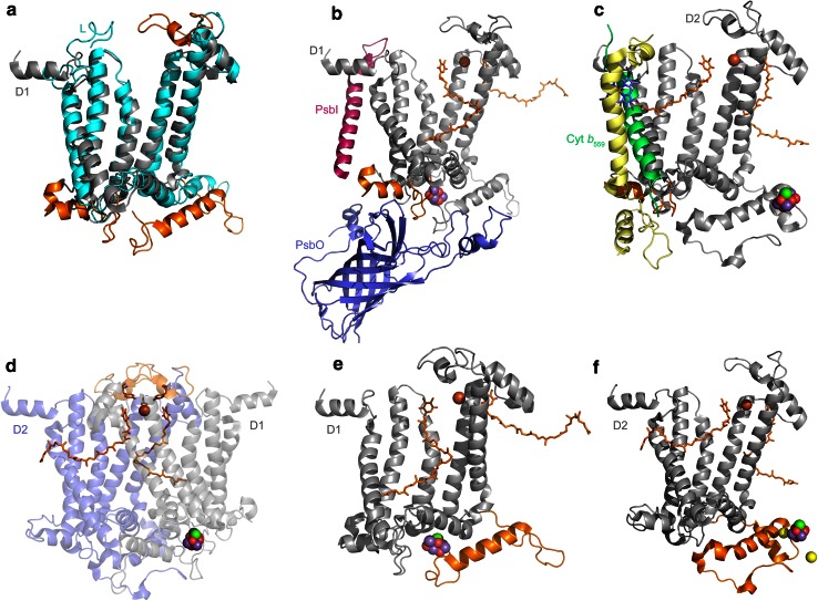
Fig. 4.
Structural comparisons of Type II reaction center proteins. a Overlap of D1 (gray) and L (cyan) subunits. Structural regions that are unique to D1 and D2 are highlighted in orange. b, c The interactions of ancillary subunits with a protein fold in D1 and D2 (orange). In D1, this region evolved to allow protein–protein interactions with the PsbI, PsbO, and CP43 subunits. In D2, it allows interactions with the Cytochrome b 559 and PsbX. The presence of this fold in D1 and D2 suggests that before the evolution of oxygenic photosynthesis, the ancestral Photosystem II was already interacting with ancillary subunits. d A unique loop (orange) only present in D1 and D2. This region contains a tyrosine that coordinates the bicarbonate ligand of the non-heme Fe2+. e, f The C-terminal extension of D1 and D2 (orange) essential for the assembly and coordination of the Mn4CaO5 cluster. This C-terminal extension contains a parallel alpha helix in both subunits, suggesting that it was present in the ancestral Photosystem II before the D1 and D2 divergence

