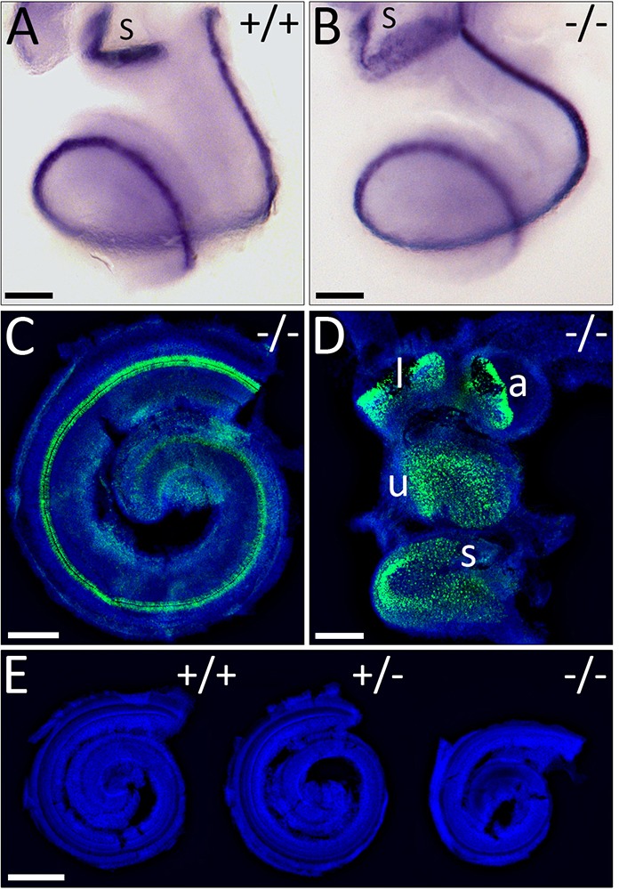
Fig. 2.

Development of cochlear and vestibular hair cells. (A,B) E16.5 wild-type and mutant cochlea revealing ATOH1 expression. (C,D) Immunolabelling of mutant cochlea and vestibule for MYO7A (green) counterstained with Hoechst to label nuclei (blue). Auditory and vestibular hair cell development is normal in WHSC1−/− mice. (E) E18.5 cochleae stained with Hoechst. Scale bars: 200 μm (A-D); 500 μm (E). l, lateral crista; a, anterior crista; u, utricle; s, saccule.
