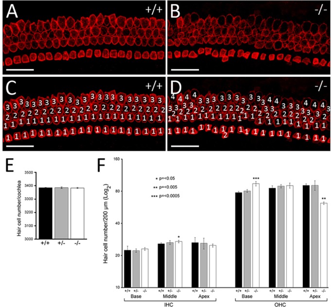
Fig. 4.

Extra hair cells in the basal and middle regions of the cochlea. (A-D) E18.5 wild-type and mutant organ of Corti immunostained for MYO7A (shown for basal turn of the cochlea). The extra rows of hair cells result from the disorganised arrangement of the hair cells. (E) Quantification of the total number of hair cells in cochleae of WHSC1+/+, WHSC1+/− and WHSC1−/− littermates showed no significant difference (n=3, P=0.4). (F) Quantification of the total number of inner hair cells (IHCs) and outer hair cells (OHCs) in a 200-μm region of base, middle and apical turns of the cochleae in heterozygous and homozygous mutants and control littermates (n=5). Note the significant increase in the number of IHCs in the middle (*P≤0.05) and OHCs in the base (***P≤0.0005), with a concomitant decrease of OHCs in the apex (**P≤0.005) of WHSC1−/− cochlea. Error bars are s.e.m. Scale bars: 25 μm.
