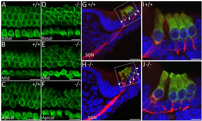
Fig. 5.

Auditory hair cells are disorganised in WHSC1−/− mice. (A-F) E18.5 wild-type and mutant organ of Corti immunostained for MYO7A (green). Differences in cell shape and size affect the arrangement of hair cells. (G-J) MYO7A and neurofilament (red) staining in sections show more spiral ganglia neuron (SGN) fibres towards outer hair cells in WHSC1−/− mice; nuclei stained with Hoechst (blue). Scale bars: 10 μm (A-F,I,J); 30 μm (G,H). (I,J) Higher magnifications of the white boxed regions in G and H, respectively. Arrowheads, hair cells; asterisk, hair cell squeezed in between other hair cells. Mid, middle region of the cochlea.
