Fig. 6.
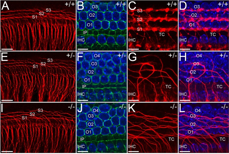
Organisation of spiral fibres is disrupted in WHSC1 mutant cochleae. Basal region of E18.5 wild-type and mutant organ of Corti immunostained for neurofilament (red), phalloidin (green) and Hoechst (blue). The innervations pattern appears normal as spiral ganglia fibres project towards their hair cell targets (A,E,I). However, whereas fibres crossing the tunnel of Corti (TC) turn basally and fasciculate to form three distinct rows in wild type (C,D), they fail to fasciculate in heterozygous and homozygous mutants (G,H,K,L). Note the orderly arrangement of similarly shaped and sized hair cells in wild-type (B) versus disorganised arrangement in mutant (F,J) cochleae. Scale bars: 30 μm (A,E,I); 10 μm (B-D,F-H,J-L). TC, tunnel of Corti; S1-S3, spiral fibre rows; O1-O4, outer hair cell rows; IHC, inner hair cells.

