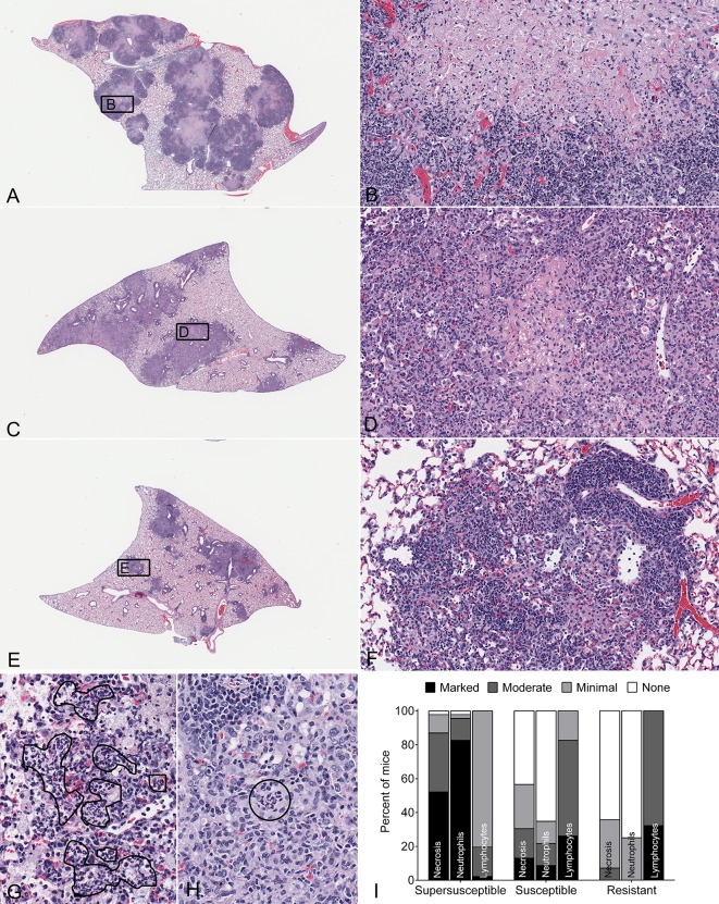
Fig. 2.
Lung lesions in M.-tuberculosis-infected DO mice. Female 8-week-old non-sibling DO mice (N=97) were infected with ∼100 M. tuberculosis (M.tb) bacilli by using an aerosol. Representative hematoxylin and eosin (H&E)-stained lung sections are shown. Supersusceptible (A) with inset showing substantial lung tissue, macrophage and neutrophil necrosis with capillary thrombosis magnified (B); susceptible (C) with inset showing a small region of necrosis surrounded by inflammatory cells that lacks thrombosis (D); resistant (E) with inset showing a small, non-necrotic lesion with an abundance of perivascular lymphocytes (F). Black lines surround neutrophils in alveolar spaces (G) and within macrophage-rich granulomas (H). Magnifications are 10× (A,C,E); 200× (B,D,F); and 400× (G,H). Two lung lobes from each mouse were scored for relative severity of each lesion type by a board-certified veterinary pathologist (G.B.) without knowledge of the groups, and the data was compiled (I).

