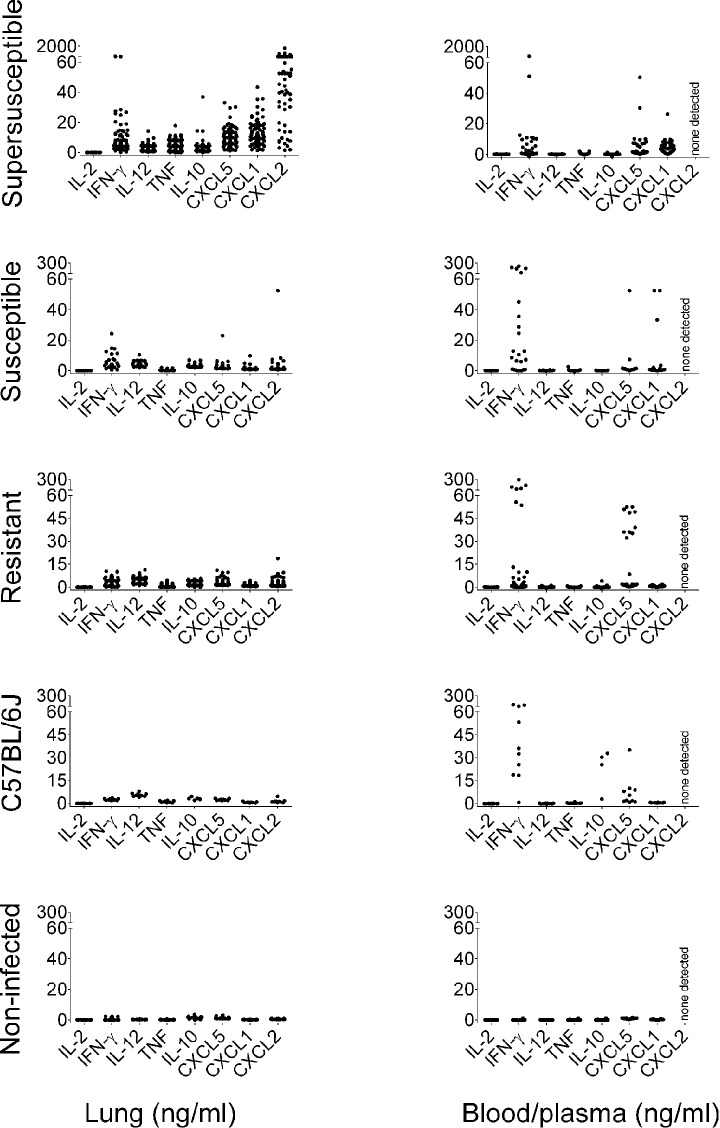
Fig. 3.
Molecular profiles of lung, and of blood and plasma in M.-tuberculosis-infected mice. Female 8-week-old non-sibling DO mice (N=166) and C57BL/6J (N=10) mice were infected with ∼100 M. tuberculosis (M.tb) bacilli by aerosol. Cytokines and chemokines were quantified in homogenized lung. Blood cytokines (IL-2, IFN-γ, IL-12, TNF, IL-10) were quantified after stimulation with antigen. Neutrophil chemokines (CXCL1, CXCL5) were quantified in plasma, but CXCL2 was not detectable. From right to left, molecular features are grouped as follows: T-cell cytokines (IL-2, IFN-γ), macrophage cytokines (IL-12, TNF, IL-10) and neutrophil chemokines (CXCL1, CXCL5, CXCL2). Each dot represents the average of duplicate or triplicate samples from one mouse. The y-axes are defined at the bottom of the figure. Data are combined from two independent experiments.

