Fig. 1.
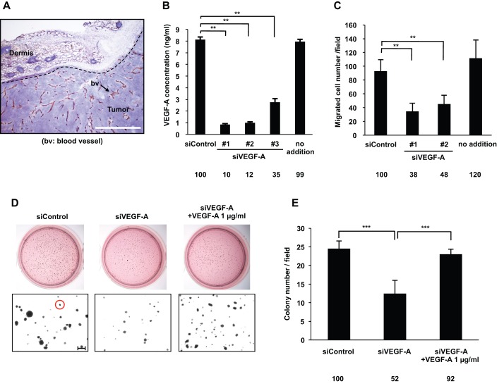
VEGF-A secreted by DJM-1 cells induced tumor angiogenesis and cancer cell proliferation. (A) Frozen sectioned DJM-1 tumors were stained with the endothelial marker CD-31 and hematoxylin. The arrow indicates blood vessels (Scale bar: 100 μm). (B) Quantification of VEGF-A concentrations secreted by DJM-1 cells. After a 72 h treatment with (20 nM, siControl, or siVEGF-A #1–3) or without siRNA (no addition), conditioned media were collected and analyzed by VEGF-A ELISA. (C) HUVEC migration assay. (B,C) Data represent the means±s.d. Percentages from the each mean relative to siControl are indicated below the graph. (D) Endogenous VEGF-A induced colony formation by cancer cells. DJM-1 cells were treated with siControl or siVEGF-A #1 (20 nM each) and seeded in soft agar. The upper panel shows the bright field of MTT staining colonies; the lower panel shows magnified colonies (Red circle: >80 μm diameter, Scale bar: 250 μm). (E) Quantitative analysis of D. The means of colony numbers in 6 fields for each condition are shown with ±s.d. Percentages from each mean relative to the siControl are indicated below the graph. **P<0.005; ***P<0.001.

