Fig. 3.
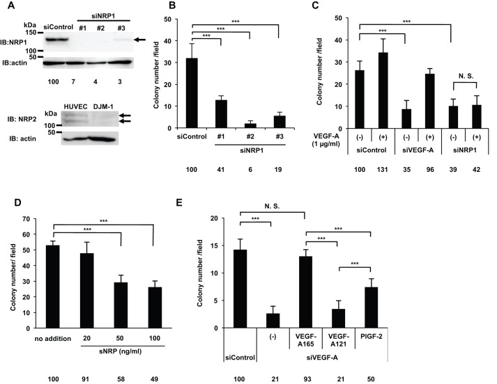
VEGF-A promoted DJM-1 cell proliferation via NRP1 in an autocrine manner. (A) A western blot shows that DJM-1 cells expressed NRP1, but not NRP2. DJM-1 cells were treated with siRNA (siControl, siNRP1 #1–3, 20 nM each) for immunoblotting the NRP1 protein (arrow indicates NRP1; 130 kDa). Percentages from each blotted protein amount relative to the siControl are indicated below each lane. Actin was immunoblotted to normalize the amounts of NRP1 (upper panel). HUVEC expressed NRP2, whereas DJM-1 cells did not (arrows indicate NRP2; 120–130 kDa, lower panel). (B) DJM-1 cell colony formation assay. Cells treated with 20 nM siControl and siNRP1 #1–3. (C) Colony formation by siVEGF-A- or siNRP1-treated DJM-1 cells. The presence or absence of exogenous VEGF-A (1 µg/ml) was indicated as (+) and (−) respectively. (D) DJM-1 cell colony formation assay in the presence of sNRP (from 20 to 100 ng/ml). (E) The graph shows the effects of VEGF-A family members (1 µg/ml each) in the siVEGF-A-treated DJM-1 cell colony formation assay. These data represent the means±s.d. Percentages from each mean relative to the siControl (B,C,E) or no addition (D) are shown below the graph. ***P<0.001; N.S., not significant.

