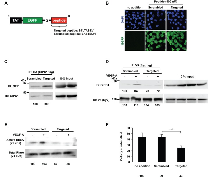
Fig. 7.
The oligopeptide that inhibited the GIPC1 and Syx interaction suppressed RhoA activity and the proliferation of DJM-1 cells. (A) A schematic of the construct that contained TAT, EGFP, and the Gly insertion prior to the Targeted peptide sequence (STLTASEV; Syx C terminus sequence). The Scrambled peptide amino acid sequence is also shown in the lower case (EASTSLVT). (B) Confirmation of the peptide incorporation into DJM-1 cells. DJM-1 cells were treated with the Scrambled or Targeted peptide (500 nM each) for 1 h. Confocal images indicated the Scrambled or Targeted peptide in the intracellular region of DJM-1 cells (green). Nuclei in the same position were shown in the upper panels (blue). Scale bar: 30 μm. (C) The co-immunoprecipitation assay with the Target peptide. HA-tagged GIPC1 was overexpressed in HEK293T cells. The Scrambled or Targeted peptide was mixed with the cell lysate and co-immunoprecipitated with GIPC1 after a 1 h rotation at 4°C. The same lysates (10% input) were immunoblotted with anti-GFP or anti-GIPC1 antibodies to normalize the amounts of the peptide and GIPC1. Percentages from each relative to the Scrambled are shown below the graph. (D) NRP1, GIPC1, and Syx vectors were transfected and expressed in HEK293T cells, which were subsequently treated with the Targeted or Scrambled peptide for 16 h. After a 10 min stimulation with (+) or without (−) VEGF-A (100 ng/ml), the cells were lysed and the indicated proteins in the cell lysates were co-immunoprecipitated with V5-tagged Syx (left panels). VEGF-A induced the GIPC1/Syx interaction in the presence of the Scrambled peptide (asterisk). On the other hand, the Targeted peptide abrogated the GIPC1/Syx interaction (asterisk). Percentages from each protein level [GIPC1 or V5 (Syx)] compared to the lane of Scrambled (−) are indicated below the lane. The same lysates (10% input) were immunoblotted with anti-GIPC1 or V5 antibodies to normalize the amounts of each protein. (E) The RhoA activity assay. DJM-1 cells were treated with the Targeted or Scrambled peptide and stimulated with (+) or without (−) VEGF-A (100 ng/ml) under anchorage-independent conditions. The same lysates (10% input) were immunoblotted with an anti-RhoA antibody to normalize the protein amounts with each treatment. Percentages from each relative to the Scrambled (−) are shown below the graph. (F) The colony formation assay. DJM-1 cells were treated with 500 nM of the Targeted or Scrambled peptide. The Targeted peptide inhibited DJM-1 cell proliferation, whereas the Scrambled peptide did not. These data represent the means±s.d. Percentages from each mean relative to the no addition control are shown below the graph. ***P<0.001.

