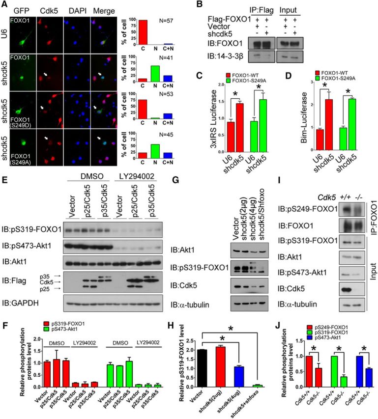
Figure 7.

Cdk5 deficiency induces nuclear accumulation of FOXO1 by inactivating AKT. A, Primary cortical neurons were transfected with control U6 or shcdk5 plasmid together with GFP-FOXO1-WT, GFP-FOXO1–S249D, or GFP-FOXO1–S249A on DIV 3, then immunostained with Cdk5 antibody on DIV 7. The subcellular localizations of GFP-FOXO1-WT, GFP-FOXO1–S249D, or GFP-FOXO1–S249A were measured in neurons displaying low Cdk5 expression; arrows indicate shcdk5 effective neurons. Representative images are shown on the left: green represents GFP; red represents Cdk5; blue represents DAPI. Quantification is shown on the right. B, Lysates of 293T cells transfected with Flag-FOXO1 together with control U6 or shcdk5 plasmid were immunoprecipitated with the Flag antibody and immunoblotted with the FOXO1 and 14-3-3β antibodies. C, D, Primary cortical neurons transfected with the GFP-FOXO1-WT or GFP-FOXO1–S249A plasmid together with the control U6 or shcdk5 plasmid plus either the 3xIRS reporter (C) or Bim reporter gene (D). Neurons were subjected to luciferase assays. Data are mean ± SEM; n = 5. *p < 0.05. E, F, N2a cells were transfected with Cdk5/p25, Cdk5/p35, or control Flag vectors. Forty-eight hours after transfection, the cells were treated with DMSO or 50 μm LY294002 for another 12 h. The lysates were immunoblotted with the indicated antibodies. F, The relative levels of pS319-FOXO1 and pS473-Akt1 were quantified. Data are mean ± SEM; n = 3. G, H, N2a cells were transfected with shRNA targeted to either Cdk5 or foxo1. Scrambled shRNA was used as a control. Forty-eight hours after transfection, the lysates were immunoblotted with the indicated antibodies. H, The relative level of pS319-FOXO1 was quantified. Data are mean ± SEM; n = 5. *p < 0.05. I, J, Brain lysates from the cortices of E16.5 Cdk5+/+ and Cdk5−/− mice were prepared, and the levels of pS249-FOXO1 were detected following immunoprecipitation with FOXO1 antibody. The other proteins were measured by directly immunoblotting with the indicated antibodies. J, The relative levels of pS249-FOXO1, pS319-FOXO1, and pS473-Akt1 were quantified. Data are mean ± SEM; n = 3. *p < 0.05.
