Figure 8.

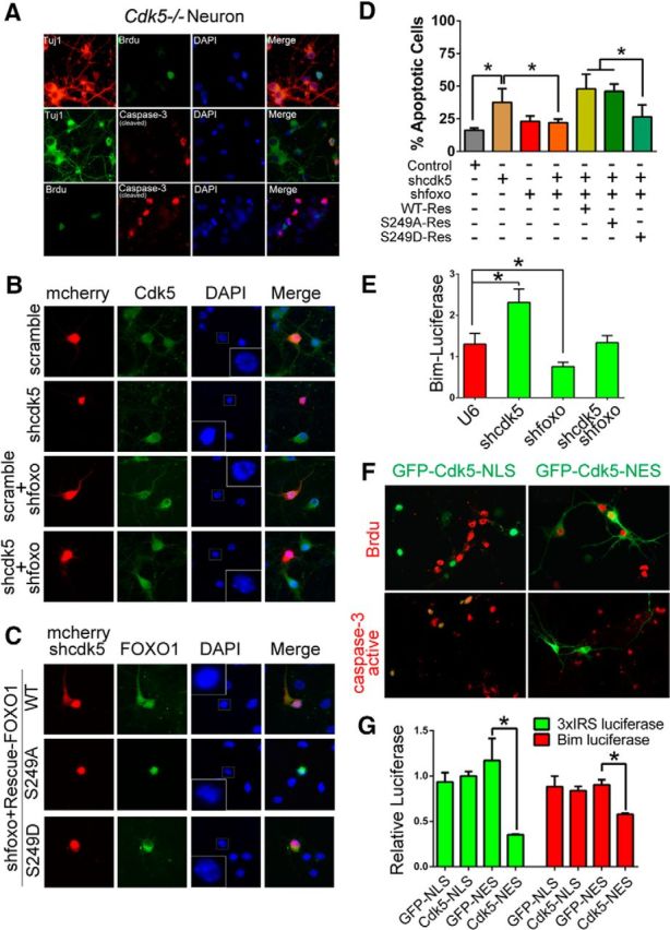
Interaction of Cdk5-dependent cell cycle and cell death activities. A, Cleaved-caspase-3 and BrdU incorporation was double stained on DIV 7 Cdk5−/− neurons. TuJ1 staining serves as neuronal cell marker. B–D, Effects of expressing mutant or wild-type FOXO1 on cortical neuronal apoptosis. B, Primary cortical neurons (DIV 3) were transfected with CMV-mCherry-IRES-U6-shcdk5 or control CMV-mCherry-IRES-U6-scramble vector either alone or together with U6-shfoxo plasmid. Cells were then immunostained with Cdk5 antibody at DIV 7. DAPI was used to counterstain nuclei to measure the neuronal death by nuclei condensation. C, Primary cortical neurons (DIV 3) were transfected with CMV-mCherry-IRES-U6-shcdk5 and shfoxo1 vector together with GFP-FOXO1-WT-Res, GFP-FOXO1–S249A-Res, or GFP-FOXO1–S249D-Res plasmid. The cells were fixed on DIV 7 and stained with DAPI to evaluate neuronal death by nuclei condensation. D, The quantification of neuronal apoptotic rate in B, C is presented in D. Data are mean ± SEM; n = 3. *p < 0.05. F, Primary cortical Cdk5−/− neurons were transfected with GFP-Cdk5-NLS or GFP-Cdk5-NES. Cleaved-caspase-3 and BrdU incorporation was detected by immunostaining. E, G, Primary cortical neurons were transfected with the indicated plasmids together with the 3xIRS-luciferase or Bim-luciferase reporter gene. The neurons were subjected to luciferase assays on DIV 7. Data are mean ± SEM; n = 5. *p < 0.05.
