. 2015 Sep 22;48(3):177–187. doi: 10.5115/acb.2015.48.3.177

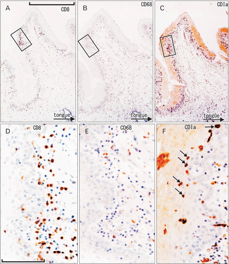
Fig. 1. CD1a-positive Langerhans cells, CD8-positive T lymphocytes, and CD68-positive macrophages in the oral floor mucosa. Immunohistochemistry of CD8 (A, D), CD68 (B, E), and CD1a (C, F). Panel (A-C) display the same mucosal fold using adjacent sections at the same magnification (scale bar in panel A=1 mm), while panels (D-F) correspond to square in panels (A-C), respectively at the same magnification (scale bar in panel D=0.1 mm). Thus, the positive cells do not show maximum density in panels (E) and (F). Some of suppressor lymphocytes have migrated into the epithelium (D), in contrast to macrophages (E). The epithelium contains abundant spherical Langerhans cells (arrows in panel F).

Fig. 1