. 2015 Sep 22;48(3):177–187. doi: 10.5115/acb.2015.48.3.177

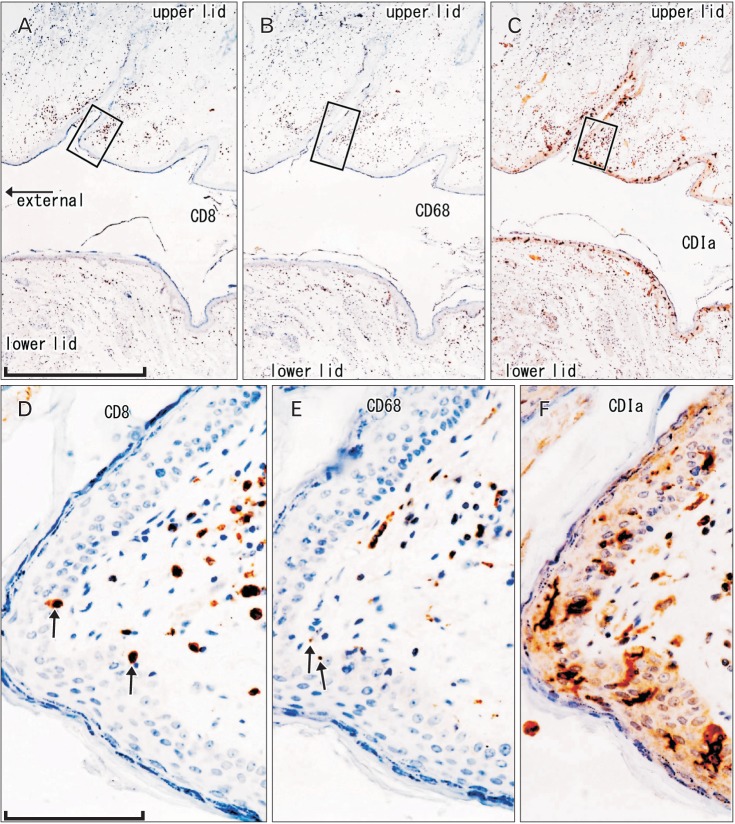
Fig. 3. CD1a-positive Langerhans cells, CD8-positive T lymphocytes, and CD68-positive macrophages in the palpebral conjunctiva. Immunohistochemistry of CD8 (A, D), CD68 (B, E), and CD1a (C, F). Panel (A-C) display the same mucosal grooves using adjacent sections at the same magnification (scale bar in panel A=1 mm), while panels (D-F) correspond to square in panels (A-C), respectively at the same magnification (scale bar in panel D=0.1 mm). Thus, the positive cells do not have maximum density (D, E). A few suppressor lymphocytes and macrophages have migrated into the epithelium (arrows in panels D and E). The epithelium contains abundant pyramidal or dendritic Langerhans cells (F).

Fig. 3