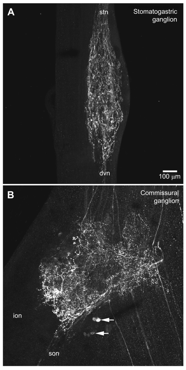
Fig. 2.

Distribution of pyrokinin-like labeling in the stomatogastric ganglion and commissural ganglion of Homarus americanus. (A) In the stomatogastric ganglion (STG), several fascicles of immunopositive axons projecting from the stomatogastric nerve (stn) arborize within the ganglion, producing a neuropile composed of a dense network of fine fibers studded with bead-like varicosities; most, if not all, of the axons producing this neuropile appear to exit the ganglion via the dorsal ventricular nerve (dvn). No pyrokinin-like immunoreactivity was seen in any of the 30 or so intrinsic STG somata. This image is a brightest pixel projection of 43 optical sections taken at approximately 2 μm intervals. (B) Pyrokinin-like labeling in the CoG consists of approximately 14 immunopositive somata (two groups indicated by arrows) and an extensive neuropile. This neuropile is likely to be derived from processes originating from both the intrinsic immunopositive cell bodies and from immunoreactive axons projecting into the ganglion from the circumoesophageal connective (coc). Immunopositive axons exiting the CoG via the superior oesophageal nerve (son) are the source of the pyrokinin labeling in the STG. No pyrokinin-like immunoreactive axons were seen exiting the CoG via the inferior oesophageal nerve (ion). This image is a brightest pixel projection of 38 optical sections taken at approximately 2 μm intervals, and is shown at the same scale as A.
