Fig. 2.
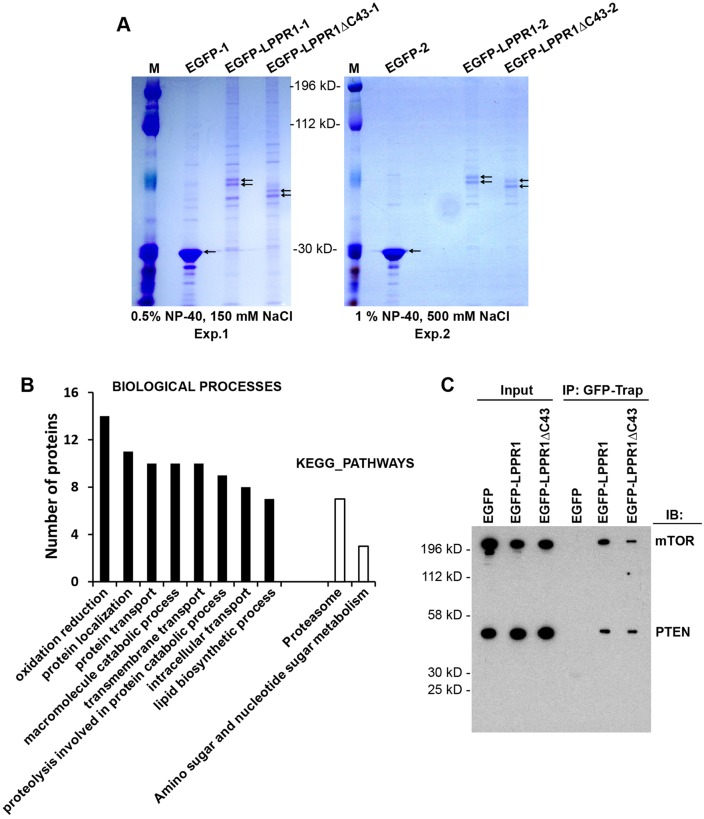
Proteome-wide identification of the LPPR1-interacting proteins. (A) Coomassie Blue staining pattern of the co-immunoprecipitated proteins. Neuro2A cells transfected with pEGFP, pEGFP-LPPR1 or pEGFP-LPPR1ΔC43 constructs were subjected to co-immunoprecipitation using GFP-Trap beads. Two independent co-immunoprecipitation experiments were performed with different detergent and the salt concentrations. M, molecular mass markers. (B) Functional annotation of the putative LPPR1-binding proteins by DAVID. Significantly enriched Gene Ontology (GO) terms for the biological process and KEGG pathways were plotted according to the number of proteins. (C) Validation of the interaction of mTOR and PTEN with LPPR1 using co-immunoprecipitation (IP). IB, immunoblot.

