Fig. 5.
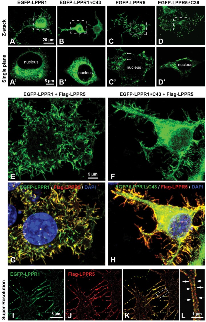
Colocalization of LPPR5 and LPPR1 on plasma membrane protrusions. (A–D) Neuro2A cells were transfected with EGFP–LPPR1, EGFP–LPPR1ΔC43, EGFP–LPPR5 or EGFP–LPPR 5ΔC39 separately. Representative z-stack confocal images collected throughout the depth of cells showing the protein distribution of EGFP–LPPR1 (A), EGFP–LPPRΔC43 (B), EGFP–LPPR5 (C) or EGFP–LPPR5ΔC39 (D). LPPR1 was expressed in endomembrane systems as well as plasma membrane protrusions, and deletion of C-terminus of LPPR1 (LPPR 1ΔC43) abolished its localization towards plasma membrane protrusions. LPPR5 was expressed mainly in plasma membrane protrusions and in some vesicle-like structures inside the cells (arrows, in C′), C-terminal truncated LPPR5 (LPPR5ΔC39) displayed similar distribution pattern as full-length LPPR5. A′–D′ are high magnification single plane images of cell body areas boxed in A–D, respectively. (E–H) Neuro2A cells were co-transfected with EGFP–LPPR1 and Flag–LPPR5, or EGFP–LPPR1ΔC43 and Flag–LPPR5. Cells were stained with anti-Flag antibody and DAPI. Co-expression of LPPR1 with LPPR5, showing LPPR1 was colocalized with LPPR5 at plasma membrane protrusions (E,G). Co-expression of LPPR1ΔC43 with LPPR5, showing colocalization of LPPR1ΔC43 with LPPR5 and that some LPPR1ΔC43 can be targeted to plasma membrane protrusions (F,H). (I–L) STED super-resolution images showing the colocalization of LPPR1 with LPPR5 on plasma membrane protrusions. The boxed area in K is highlighted in L, showing a strong colocalization observed along the two opposed membranes of the filopodia (arrows).

