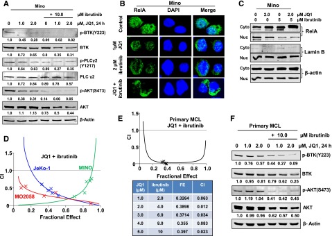
Figure 4.

Compared with treatment with either agent alone, combined treatment with JQ1 and ibrutinib exerts synergistic lethal activity against cultured and primary MCL cells. (A) Representative immunoblots from Mino cells treated with JQ1 and/or ibrutinib, as indicated, for 24 hours. The numbers beneath the bands represent densitometry analysis performed on the blots and normalized to the β-actin loading control. (B) Mino cells were treated with JQ1 and/or ibrutinib for 24 hours. Confocal immunofluorescence microscopy was performed for RelA subcellular localization. Nuclei were stained with DAPI. Original magnification, ×63. (C) Nuclear and cytosolic fractions were prepared from Mino cells treated as indicated for 24 hours and the expression levels of RelA in each fraction were determined by immunoblot analyses. The localization of Lamin B served as a fraction and loading control. (D) MO2058, JeKo-1, and Mino cells were treated with JQ1 and ibrutinib at a constant ratio for 48 hours. The percent of apoptotic cells was determined by flow cytometry. Median dose effect and isobologram analyses were performed utilizing CalcuSyn. CI values <1.0 indicates a synergistic interaction of the two agents in the combination. Doses of drugs, fractional effect, and CI values are provided in supplemental Figure 6A. (E) Primary MCL cells were treated with JQ1 and ibrutinib at a constant ratio for 48 hours. The percent of nonviable cells was determined by flow cytometry. Median dose effect and isobologram analyses were performed utilizing CalcuSyn. CI values <1.0 indicates a synergistic interaction of the two agents in the combination. (F) Primary MCL cells were treated with the indicated concentrations of JQ1 and/or ibrutinib for 24 hours. Then, total cell lysates were prepared and immunoblot analyses were conducted as indicated. The numbers beneath the bands represent densitometry analysis performed on the blots and normalized to the β-actin loading control. Cyto, cytoplasmic; DAPI, 4′,6 diamidino-2-phenylindole; FE, fractional effect; Nuc, nuclear.
