


Fig. 4.
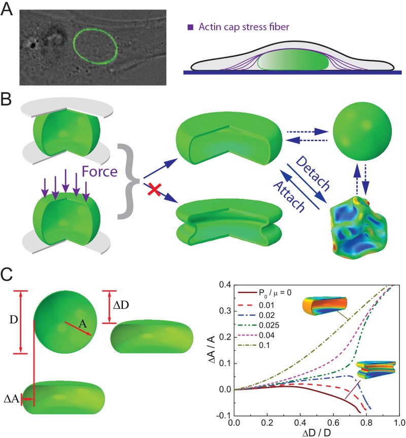
Nuclei in adherent cells. (A) The nucleus in a GFP–lamin-A-transfected MEF attached to a collagen-coated glass-bottomed dish. The nucleus is compressed by perinuclear actin stress fibers from above. (B) In our model, forces from the perinuclear actin fibers are modeled as a compressive plate. The surrounding microtubules are modeled by a positive mechanical pressure on the nucleus. Our FEM simulations show that in order to obtain the nuclear shape observed in attached cells, the nucleus has to be ‘inflated’ by a negative pressure; otherwise, the nuclear envelope will buckle and invaginate. As the cell detaches, owing to a large cell volume reduction, the osmotic pressure outside the nucleus becomes larger and the pressure difference becomes positive. The nucleus responds to the changing pressure by developing surface wrinkles. (C) Left, in the adhered state, the nucleus is compressed by actin stress fibers and resembles a pancake. In the deformed state the change in nuclear radius and height are denoted as ΔA and ΔD, respectively. Right, the computed shape of the nucleus in the adherent state. The pressure difference, P0/μ, must be sufficiently large such that the nuclear envelope will not buckle under actin force, where μ is the shear modulus of the nuclear envelope.