


Fig. 6.
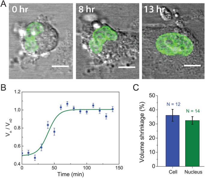
Nuclear volume change is reversible and consistent with the osmotic shock results. (A) The nuclear shape change is reversible. A GFP–H2B-histone-transfected MEF was initially treated with trypsin. After the cell had detached partially, it was gently washed with pure medium and cell became fully attached. In this process, the nucleus transformed from a wrinkled morphology to a round shape, reversing the shape changes seen in Fig. 2. Scale bars: 10 μm. (B) Volume change in the nucleus of a MEF cell during cell going from the detached state to the attached state (reverse of Fig. 2, see images in A). Vn0 is the nuclear volume in the adherent state. Vn denotes the nuclear volume during detachment. Results are mean±s.e.m., n=31 cells (>10 cells per trial, as indicated in the figure, repeated three times). (C) Volume reduction after hypertonic shock on adherent cells. A similar volume shrinkage occurred in the cell and the nucleus. Therefore, extracellular osmotic content changes cytoplasmic osmotic pressure, which drives nuclear volume shrinkage.