Fig. 3.

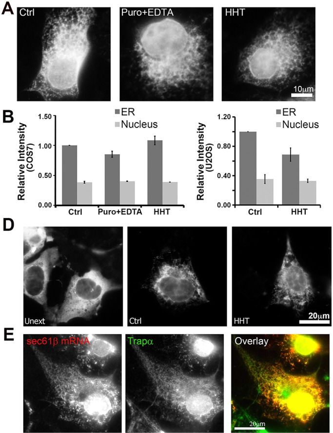
ER association of overexpressed GFP–Sec61b mRNA is partially independent of translation. (A,B) COS7 and U2OS (C,D) cells were transfected with plasmid encoding GFP–Sec61b and allowed to express mRNA for 18–24 h. Cells were then treated with DMSO (Ctrl), puromycin (Puro) or homoharringtonin (HHT) for 30 min, and then extracted with digitonin with or without EDTA. Cells were then fixed and stained for mRNAs using a specific FISH probe against the GFP-coding sequence. Cells were imaged (A,D), and the fluorescent intensities were quantified (B,C). To control for changes in staining, nuclear fluorescent intensities were also analyzed. Each bar represents the mean±s.e.m. of three independent experiments, with each experiment consisting of at least 30 cells. (E) U2OS cells expressing GFP–Sec61b were treated with HHT and then digitonin extracted. Cells were then stained for the GFP–Sec61b mRNA, and immunostained with the ER marker Trapα. Images in E are from a single field of view including a color overlay showing the GFP–Sec61b mRNA in red and Trapα in green. Scale bars: 20 µm.
