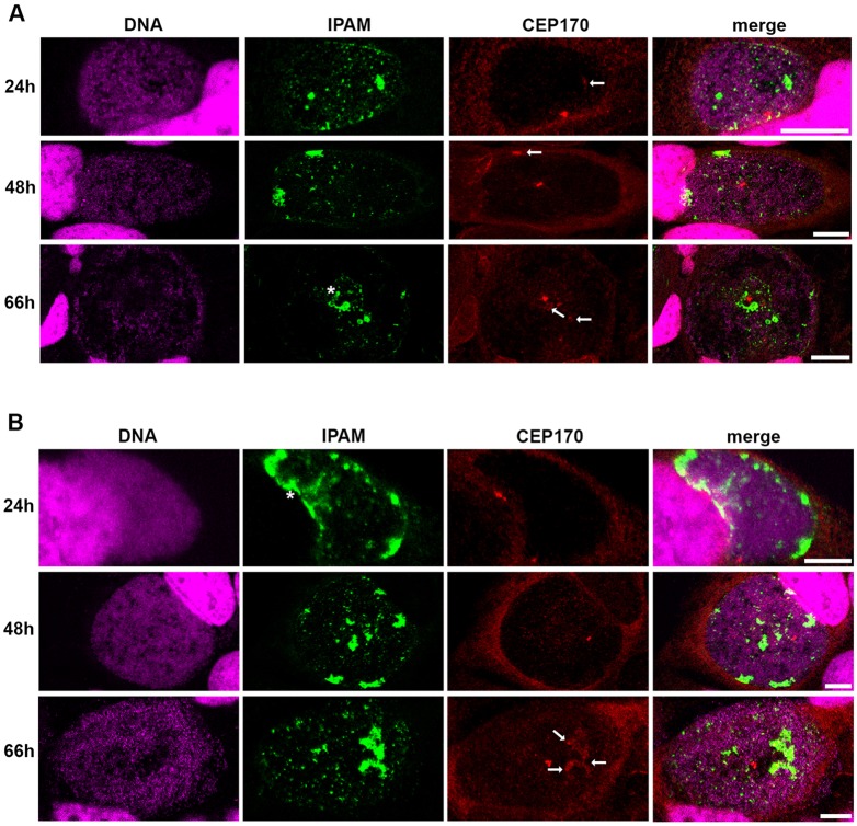
Fig. 5.
IPAM and CEP170 location in C. trachomatis-infected cells. (A,B) HeLa cells (A) or U2OS cells (B) were infected with C. trachomatis for 24 h, 48 h or 66 h and labeled for DNA (magenta), endogenous IPAM (green) and CEP170 (red). Asterisk indicates IPAM patches (as described in supplementary material Fig. S2A) in proximity of CEP170 dots. Arrows indicate diffuse CEP170 signal associated to IPAM. Scale bars: 10 µm.

