Fig. 6.
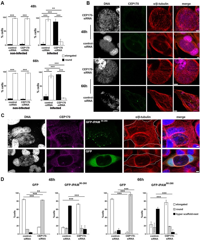
CEP170-IPAM interaction is essential to maintain cell shape and MT organization in Chlamydia-infected cells. (A) HeLa cells were transfected with control-siRNA or CEP170-siRNA, and infected with C. trachomatis where appropriate. Cell shape was assessed and categorized after 48 h and 66 h. Cells in mitosis identified by DNA staining were excluded. **P<0.01, ***P<0.005. (B) HeLa cells transfected with CEP170-siRNA or control-siRNA were infected with C. trachomatis and fixed after 48 h or 66 h. Cells were stained for DNA (grey), CEP170 (green) and α/β-tubulin (red). Panels show maximum projections of four z-sections. Scale bars: 10 µm. (C) HeLa cells were transfected with CEP170-siRNA or control-siRNA for 24 h, prior to infection with C. trachomatis and transfection with plasmid encoding GFP or GFP-IPAM90-268 (green). Cells were fixed 48 or 66 h later and labeled for DNA (gray, blue in merged panel) CEP170 (purple) and α/β-tubulin (red). Panels represent maximum projections of four z-sections in cells 48 h after infection, showing a representative ‘hyper scaffold-nest’. The cell periphery is outlined in gray. Scale bars: 5 µm. (D) Following confocal imaging, cells shape was categorized (as described in supplementary material Fig. S2E) for C. trachomatis-infected cells. ***P<0.005, **P<0.01.

