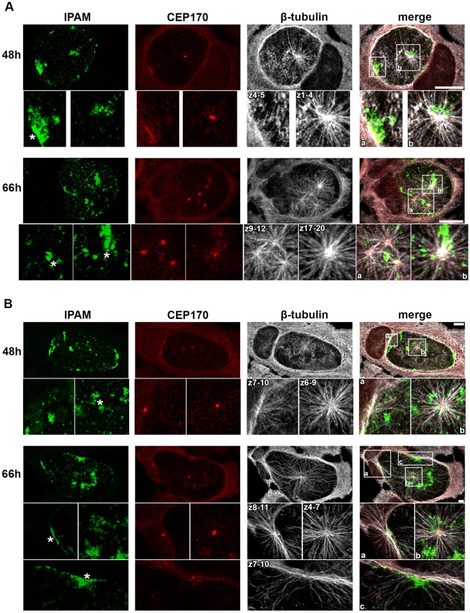
Fig. 8.
Endogenous IPAM localizes to sites of MT assembly in C. trachomatis-infected cells. (A,B) HeLa cells (A) or U2OS cells (B) were infected with C. trachomatis for 48 h or 66 h prior to MT regrowth for 5 min. Cells were fixed and labeled for endogenous IPAM (green), CEP170 (red) and β-tubulin (gray). Upper panels show maximum projections through the entire cell volume. Lower panels, show the maximum projection of the indicated z-section from the marked region of interest. Asterisks indicate the IPAM patches where MTs regrow. Scale bars: 10 µm.

