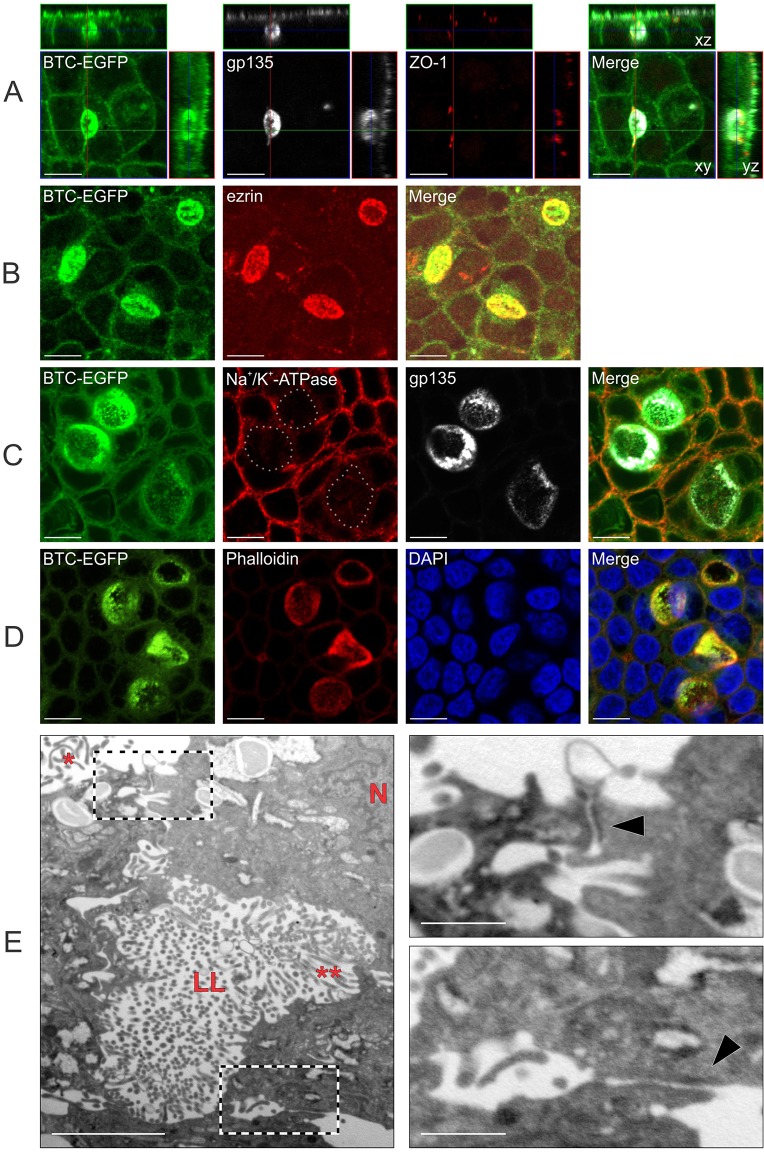
Fig. 4.
BTC mistrafficking results in lateral lumen formation in polarized MDCK cells. (A–D) Polarized MDCK cells stably expressing (C3/TM)BTC–EGFP were fixed and stained with polarity markers. In A, immunofluorescence for gp135 (white) and ZO-1 (red) is shown with BTC–EGFP fluorescence (green). Confocal xy projections for each stain are shown in the center with xz and yz projections displayed to the top and right, respectively. In B, cells were stained for an apical marker, ezrin (red). In C, cells were stained for a basolateral marker, Na+/K+-transporting ATPase α1 subunit (red), and an apical marker, gp135 (white); in the second panel, lateral lumen membranes are highlighted with dotted lines. In D, cells were stained for Rhodamine–phalloidin (red) and for nuclei with DAPI (blue). (E) Polarized MDCK cells stably expressing (C3/TM)BTC–EGFP were fixed and processed for transmission electron microscopy analyses, as described in Materials and Methods. The main figure on the left shows lateral lumen (LL) with microvilli (**). Apical microvilli and nuclei are indicated with * and N, respectively. On the right, high-magnification views of the two boxed areas from the left panel show tight junctions (black arrowheads) above (top panel) and below (bottom panel) the lateral lumen. Scale bars: 10 µm (A–D); 2 µm (E, main image); 0.5 µm (E, insets).

