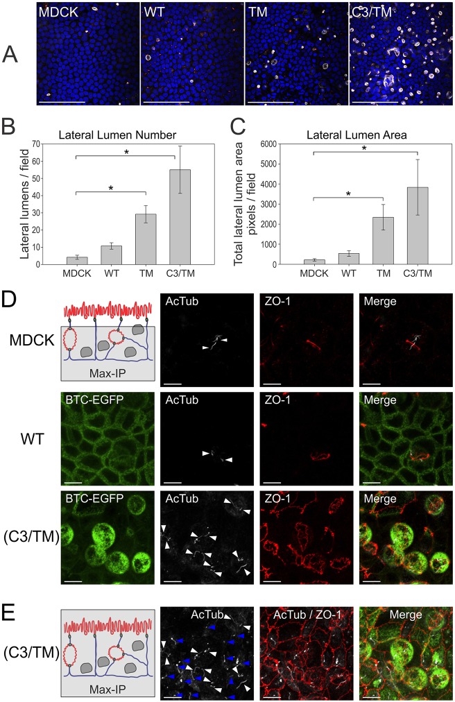
Fig. 6.
Detection of lateral lumens in polarized MDCK cells – enhancement by BTC mistrafficking. (A) Parental MDCK cells and MDCK cells stably expressing the indicated BTC constructs were polarized on Transwell filters, and then fixed and stained with DAPI (blue), and for gp135 (white) and ZO-1 (red). Quantification of number (B) and area (C) of lateral lumens. *Statistically significant difference, P<0.05. See Materials and Methods for details. (D) Polarized MDCK cells expressing the indicated constructs were fixed and stained for acetylated tubulin (AcTub, white) and ZO-1 (red). Maximum intensity projections (Max-IP) of the z-stacks, excluding the apical surface, are displayed; the area included in the analysis is shaded in gray in the schematic at the top left. Individual primary cilia are indicated by white arrowheads. (E) Maximum intensity projections that include the apical surface at the apex (schematic on left) from the corresponding area imaged in D for (C3/TM)BTC–EGFP-expressing MDCK cultures are displayed. White arrowheads indicate primary cilia within the lateral lumens; blue arrowheads indicate apical primary cilia at the apex. In the schematics in D,E: red, apical surfaces; blue, basolateral membranes. WT, wild type. Scale bars: 100 µm (A); 10 µm (D,E).

