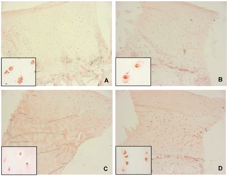
Figure 1. BMP-2 immunohistochemistry at different stages of osteoarthritis.

A: normal cartilage (ICRS: 0), B-D: osteoarthritic cartilage with weak (B, ICRS: 1), moderate (C, ICRS: 2) and severely (D, ICRS: 3) damage; all sections show intracellular localization of BMP-2 throughout all cartilage layers, extra cellular BMP-2 could only be observed in osteoarthritic cartilage (C, D), original magnification: 2.5x, inserts: 63x.
