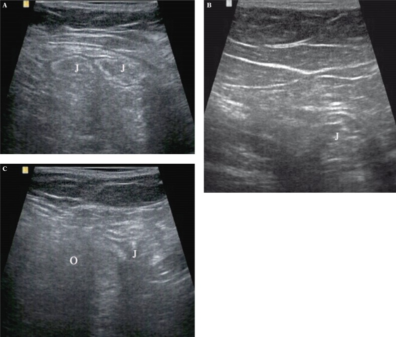
Fig. 6.
In 62 years old woman bilateral Spigelian hernias were visualized: A – greater on the right side contains small intestine loop (J); B – smaller on the left side with small fragment of small intestine (J); C – the same hernia as in fig. 6 A decreased under the compression of the transducer. In this figure small fragment of intestine (J) and greater omentum (O) are visible

