Fig. 1.

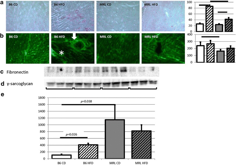
The HFD B6 hearts present with fibrosis, which is absent in the HFD MRL hearts. a Picro Sirius Red staining was enhanced in the HFD B6 hearts compared to the other three groups. Initial magnification ×100. Histogram of ImageJ quantification follows, N = 3, bar represents p < 0.05. b Representative immunofluorescent microscopy of fibronectin stained tissues confirms the increased fibrosis in HFD B6 hearts. Initial magnification ×100. Histogram of ImageJ quantification follows, N = 3, bar represents p < 0.05. c Immunoblot of fibronectin verifies increased fibrosis in the HFD B6. d Control γ-sarcoglycan immunoblot. e Quantification (N = 6, B6 CD versus B6 HFD p = 0.026, B6 CD versus MRL CD p = 0.038)
