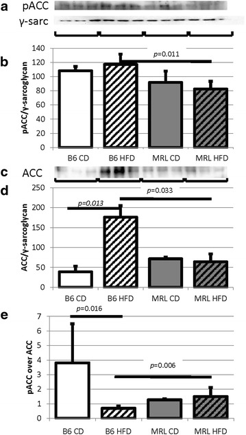
Fig. 6.

Decreased MRL cardiac ACC indicates increased fatty acid metabolism. a pACC immunoblot. b Quantification normalized to γ-sarcoglycan shows the MRL tissues have reduced pACC (N = 6, B6 HFD versus MRL HFD p = 0.011). c ACC immunoblot. d Quantification normalized to γ-sarcoglycan shows the MRL tissues have reduced ACC (N = 6, B6 HFD versus MRL HFD p = 0.033, B6 CD versus B6 HFD p = 0.013)
