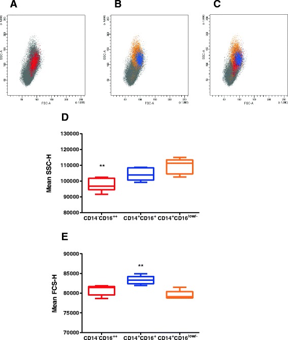
Figure 3.

Size and granularity of bovine myeloid sub-populations. Live single PBMC were gated as shown in Figure 2C and each highlighted in different colours for identification in side and forward scatter plots. The size and granularity of the populations was determined by their characteristic side and forward scatter plots: in red, CD14−CD16++ (A); in orange, CD14+CD16low/- and, superimposed, the CD14+CD16+ population in blue (B). Combining the plots (C) showed the difference of size and granularity of these cells. Data shown are for one representative animal out of six. (D, E) Box and whiskers plots for the arithmetic mean SSC-H (D) and FSC-H (E) for each myeloid subset (n = 6) are presented as the mean values ± SE, showing the difference in size and granularity of the populations. ** denotes significant difference (p < 0.001).
