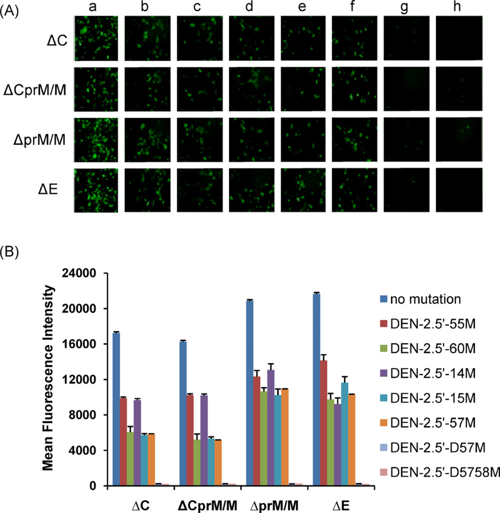
Fig. 5.
(A) Fluorescent images of Δ-DENV/GFP plasmid-transfected cells at 48 h pt. Δ-DENV/GFP replicons with (a) wt 5′NCR, (b) DENV-2.5′-55 M, (c) DENV-2.5′-60M, (d) DENV-2.5′-14M, (e) DENV-2.5′-15M, (f) DENV-2.5′-57M, (g) DENV-2.5′-D57M, and (h) DENV-2.5′-5758M. (B) MFI of GFP in BHK-21 cells transfected with Δ-DENV/GFP plasmids at 48 h pt. MFI in mutant-transfected cells were all significantly less (P < 0.001) than those in cells transfected with corresponding replicon containing wt 5′NCR.

