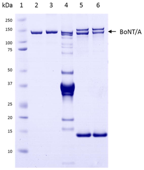
Figure 1.
1D gel image of the BoNT/A toxin samples. Lane 1: Molecular weight marker; Lane 2 and 3: purified BoNT/A1 and /A2. Lane 4: BoNT/A5 complex; Lane 5 and 6: A1 and A5 after extraction by toxin specific monoclonal antibody attached to magnetic beads. Bands at approximately 15 kDa and 155 kDa are associated with the antibody-coated streptavidin beads.

