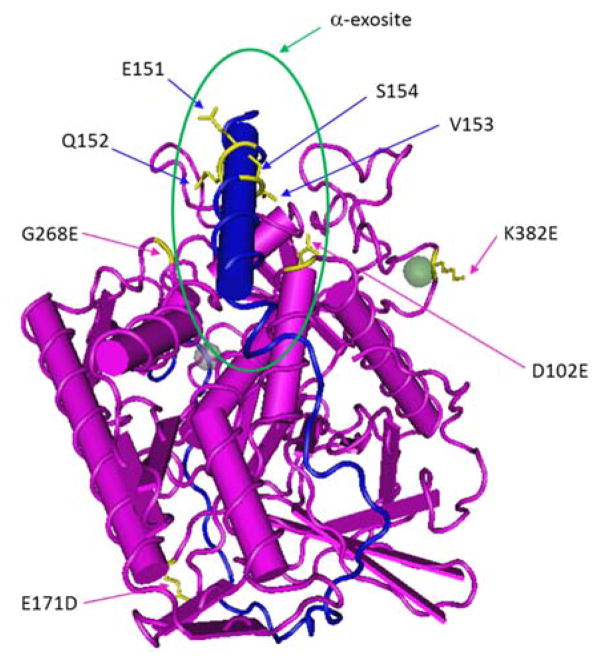
Figure 4.
Complex structure of the inactive light chain of BoNT/A1 (pink) and SNAP-25141–204 (blue)(Breidenbach and Brunger 2004). The residues differentiated in BoNT/A5 and some selected residues in the peptide substrate are colored as yellow. Green circle represents the approximate location of α-exosite.

