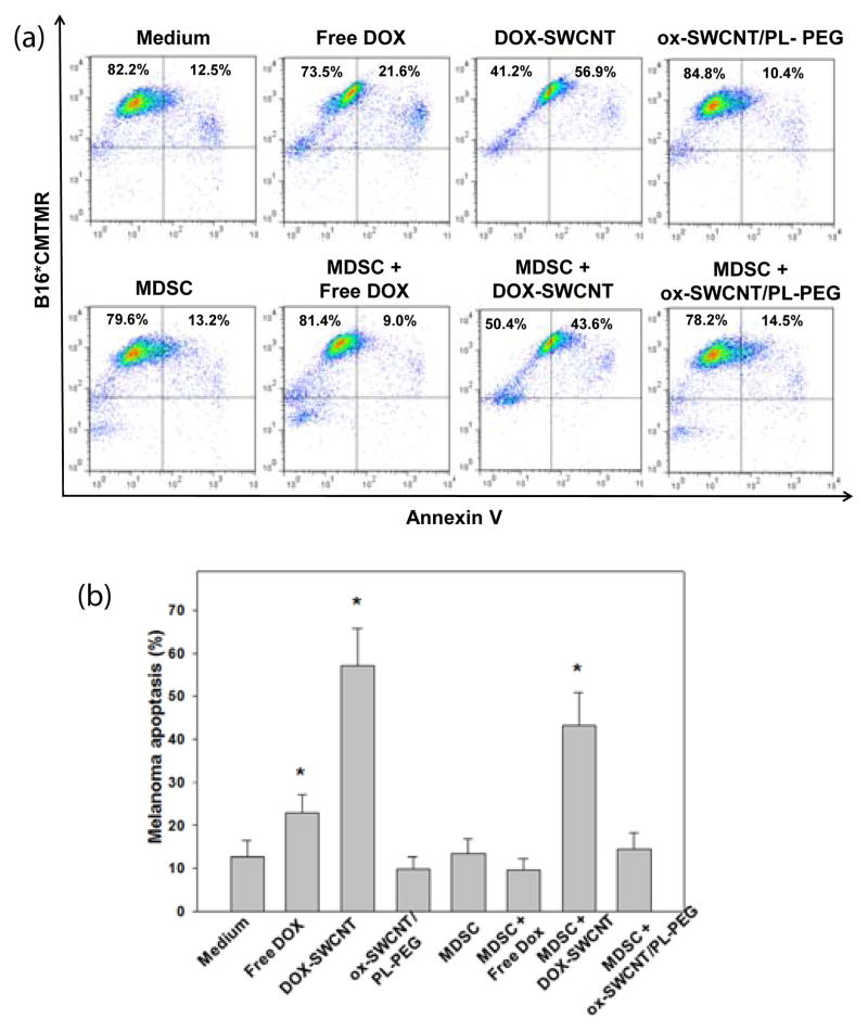
Fig. 5. Cytotoxic effects of free DOX vs. DOX-SWCNT in B16 melanoma cells and bone marrow-derived, tumor-activated MDSC.
DOX-SWCNT, but not free DOX, induces significant apoptosis of B16 melanoma cells even in the presence of MDSC. B16 melanoma cells and bone marrow-derived tumour-activated MDSC were generated as described in Supporting Information and co-cultured in the presence of free DOX or DOX-SWCNT. ox-SWCNT/PL-PEG served as a control. The level of tumour cell apoptosis was determined 24 h later by Annexin V binding as described in Supporting Information. All cell cultures were set in triplicates, and results are shown as representative flow cytometry dot plots in (a) and the mean ± SEM (standard error of the mean) (N=3) in (b).*, p<0.01 versus control (medium) group (One way ANOVA).

