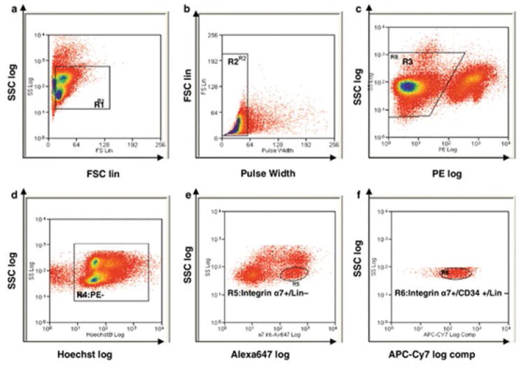
Figure 3. Example methodology for FACS purification of satellite cells (adapted from Pasut, et al. 2012 [53]).
Dot plots representing the sequential gating strategy used to identify satellite cells from a heterogonous muscle sample based on: (a) Side Scatter (SSC) and Forward Scatter (FSC), (b) Doublets discrimination, (c) PE- gating to remove CD45 and CD11b blood lineage cells, Sca-1 mesenchymal progenitors, and CD31 endothelial cells, (d) Hoechst staining for live (Hoechst+) and dead (Hoechst−) cells, (e) Integrin α7+ and Sca-1- (Lin−) gating, and finally (f) Integrin α7+ and CD34+ gating to sort the population defined as satellite cells. As the plots above illustrate, unique populations are rarely completely distinct, and substantial technical expertise is necessary to design such gating strategies properly.

