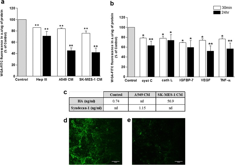
Fig. 5.

Exposure of lung tumour CM and secreted-proteins significantly altered the brain endothelial glycocalyx. a and b Integrity of the hCMEC/D3 glycocalyx was assessed by a CBF assay following exposure of ECs for 30 min or 24 h to A549 and SKMES-1 CM (a), or 80 ng/ml CC, 10 ng/ml CL, 200 ng/ml IGFBP-7, 0.2 ng/ml VEGF and 160 pg/ml TNF-α (b). Data from 6 independent experiments, carried out in sextuplicate, is presented as WGA-FITC fluorescence/μg protein and these values were calculated as a percentage of control. *p ≤ 0.05, **p ≤ 0.01 vs control level. c Hyaluronan (HA) and syndecan-1 levels were measured by ELISA in hCMEC/D3 growth medium following 30 min treatment with fresh DMEM-BS (control), A549 or SK-MES-1 CM. SK-MES-1 CM vs control = p = 0.007, n = 6. nd = not detected. d and c Representative confocal images of FITC-WGA stained hCMEC/D3 cells before (d) and (e) after 30 min exposure to SKMES-1 CM. Scale bar = 40 μM
