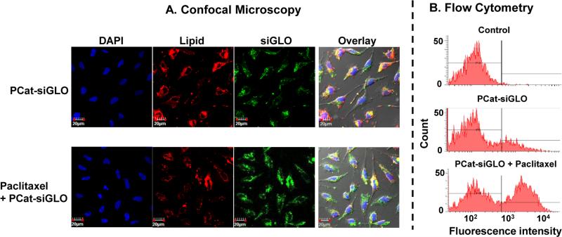
Figure 1. Paclitaxel enhanced the cytoplasmic release of siGLO from PCat-siRNA lipoplex and/or endosome/lysosome.
Cells were incubated with PCat-siGLO, with or without paclitaxel (2 nM) cotreatment. Cell nuclei were stained with DAPI (blue). PCat, which contained rhodamine-labeled DOPE, showed red fluorescence. siGLO showed green fluorescence. siGLO contains a nucleus translocation sequence and, upon release from lipoplex siRNA and/or endosomes, enters the nucleus. (A) Confocal microscopy results. Colocalized red and green signals indicate intact PCat-siGLO. Separate green signals indicate siGLO dissociated from the lipoplex. (B) Flow cytometry analysis of siGLO-containing nuclei. The background fluorescence for flow cytometry gating was established using untreated controls (indicated by vertical line). The fraction of nuclei with fluorescence signals above the background fluorescence (indicated by horizontal line) was determined.

