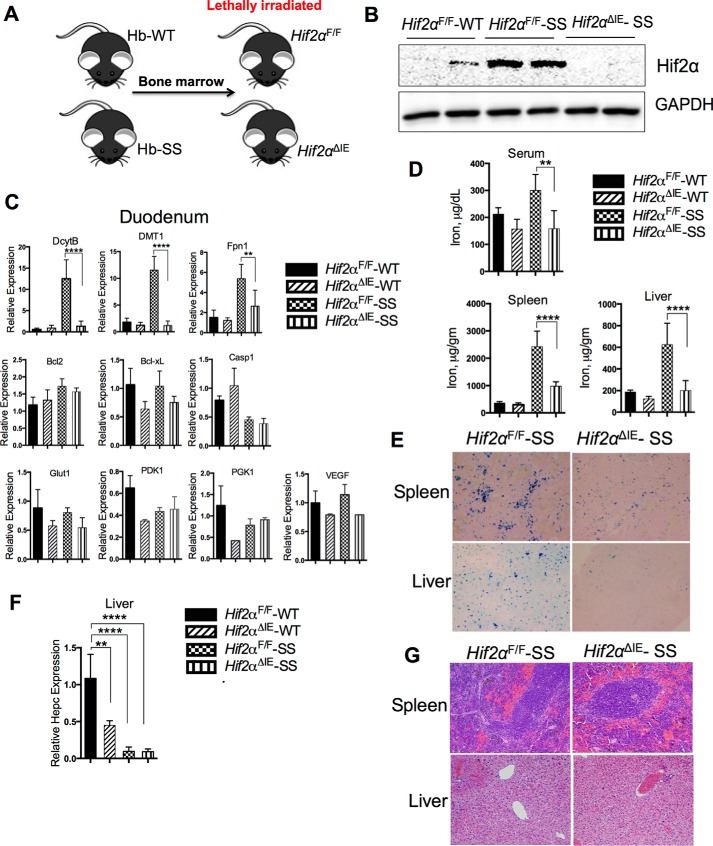
FIGURE 1.
Disruption of intestinal HIF-2α improves SCD-induced tissue iron overload. A–C, Hif2αF/F and Hif2αΔIE mice were lethally irradiated and transplanted with Hb-WT or Hb-SS bone marrow (A) and analyzed 6 months later for HIF-2α protein expression in the duodenal mucosa (B) and gene expression analysis of duodenal Dcytb, Dmt1, Fpn1, Bcl2, Bcl-xL, Casp1, Glut1, PDK1, PGK1, and VEGF1 (normalized to β-actin) (C). D, serum (μg/dl) and tissue iron content (calculated as μg per gm of wet weight). E, Perls' iron stain of the spleen and liver. F, gene expression analysis of liver hepcidin (Hepc) normalized to β-actin. G, H&E staining of spleen and liver. Bar graph represents the mean value ±S.D. ****, p < 0.0001; **, p < 0.01; *, p < 0.05; ns, not significant.

