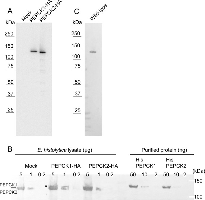
FIGURE 3.
WB analysis of EhPEPCKs with anti-HA (A) and anti-EhPEPCK (B and C) antibodies. A, expression of EhPEPCK1-HA and EhPEPCK2-HA was confirmed. Note that proteins with the expected size (∼130 kDa) were expressed. Amebic lysates containing 5 μg of protein were applied per lane on SDS-PAGE. B, expression of EhPEPCKs in EhPEPCK1-HA- or EhPEPCK2-HA-expressing and mock control E. histolytica transformants. Total lysates of the above mentioned transformants (0.2–5 μg/lane) and purified histidine-tagged PEPCK1 and -2 (2–50 ng/lane) were electrophoresed on SDS-PAGE and subjected to WB analysis with anti-EhPEPCK antiserum. Note that lysates from mock transformant showed two bands, whereas EhPEPCK1-HA-expressing transformant showed one additional band (shown with an asterisk) corresponding to PEPCK1 with the HA tag. Recombinant PEPCKs possess the His tag at the N terminus and were purified to homogeneity using Ni-NTA and MonoQ columns. C, specificity of anti-EhPEPCK antibody. Approximately 1 μg of lysate from the wild-type E. histolytica was reacted with anti-EhPEPCK antibody.

