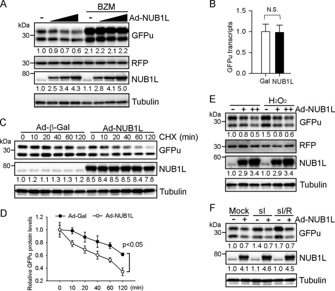
FIGURE 5.
NUB1L overexpression enhances the degradation of GFPu. NRVMs were first infected with Ad expressing GFPu (Ad-GFPu) and RFP (Ad-RFP) for 24 h and subsequently with Ad-NUB1L or Ad-Gal. A, representative Western blots of total cell extracts are shown. Ad-NUB1L was infected at 5, 10, and 20 MOI and Ad-Gal at 20 MOI. BZM (100 nm) was added 6 h prior to cell harvesting. B, quantitative real-time PCR analysis of GFPu transcripts. N.S., not significant. C and D, CHX chase assay. Representative Western blots (C) and the quantification (D) are shown. GFPu protein levels at 0 h were set as 1. E and F, cells were treated with H2O2 (100 μm) for 8 h (E), sI for 6 h, or sI followed by reperfusion for another 6 h (sI/R) (F) prior to cell harvesting. Ad-NUB1L was infected at 10 and 20 (E) or 10 MOI (F). Representative Western blots of total cell extracts are shown. The fast-migrating band of GFPu is its N-terminal truncated form. The densitometries of GFPu normalized to RFP or NUB1L are denoted in A, C, E, and F.

