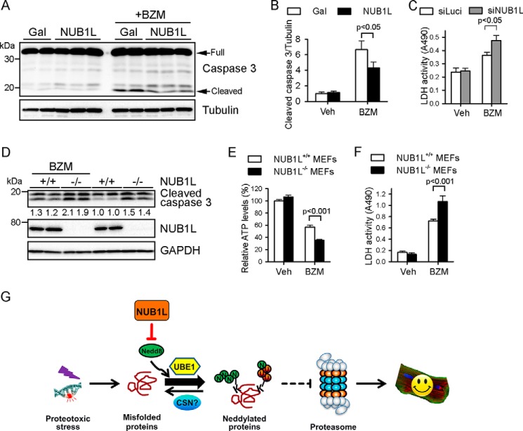
FIGURE 9.
NUB1L ameliorates proteotoxic stress-induced cytotoxicity. A–C, NRVMs were infected with the indicated adenovirus (A and B) or transfected with the indicated siRNA (C) for 48 h before being subjected to BZM (100 nm) treatment for another 24 h. Representative Western blots of total cell extracts (A) and the densitometric analysis (B) are shown. n = 4–6/group. Veh, vehicle. C, culture medium was collected for the lactate dehydrogenase (LDH) activity assay. D–F, NUB1L wild-type (+/+) or deficient (−/−) MEFs were treated with BZM (100 nm) for 24 h. D, representative Western blots of total cell lysates and the densitometries of cleaved caspase 3 relative to those of GAPDH. E, cell viability assay by measuring cellular ATP levels. F, LDH activity assay. For cell viability and LDH release assays (C, E, and F), data are representative of three independent experiments. n = 6–8 samples/group for each replication. G, model of the function of NUB1L in proteasomal proteolysis.

