FIGURE 3.

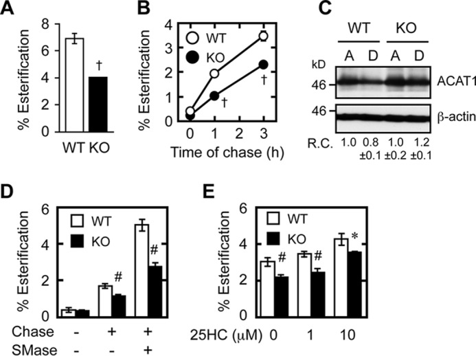
Retrograde cholesterol transport is impaired in Abca1−/− cells. A, esterification of [3H]cholesterol at steady-state level. MEFs were set up as described in Fig. 1. On day 2, cells were labeled with [3H]cholesterol for 8 h in medium A. After washing [3H]cholesterol present in the medium, cells were incubated in medium A for 16 h for equilibration, and further incubated for 6 h in medium F. [3H]Cholesterol and [3H]CE in cells were analyzed. †, p < 0.001 (n = 3). B, esterification of cell surface cholesterol. MEFs were set up as described in Fig. 1. On day 3, cells were pulse-labeled with [3H]cholesterol for 30 min in medium B (medium containing 0.1% BSA) at 37 °C, washed twice, and chased in medium F (serum-free medium) for various times as indicated. [3H]Cholesterol and [3H]CE in cells were analyzed. †, p ≤ 0.001 (n = 3). C, ACAT1 protein expression. MEFs were incubated in medium A or D for 2 days. ACAT1 and β-actin expressions were analyzed by immunoblots. Relative changes in ACAT1 expression (ACAT1/β-actin) (R.C.) are indicated at the bottom. D, effect of SMase on retrograde cholesterol transport. MEFs were set up and pulse-labeled with [3H]cholesterol as in B and chased or not chased for 1 h with or without SMase (0.1 unit/ml). Cellular [3H]cholesterol and [3H]CE were analyzed. #, p < 0.003 (n = 3). E, effect of 25HC on the esterification of PM cholesterol. MEFs were set up and pulse-labeled with [3H]cholesterol as in B and chased or not chased for 3 h with or without 25HC (1 or 10 μm as indicated). Cellular [3H]cholesterol and [3H]CE were analyzed. *, p < 0.05; #, p < 0.01 (n = 3). Error bars represent S.D. Statistical analyses were performed by one-way ANOVA with Tukey-Kramer post hoc test.
