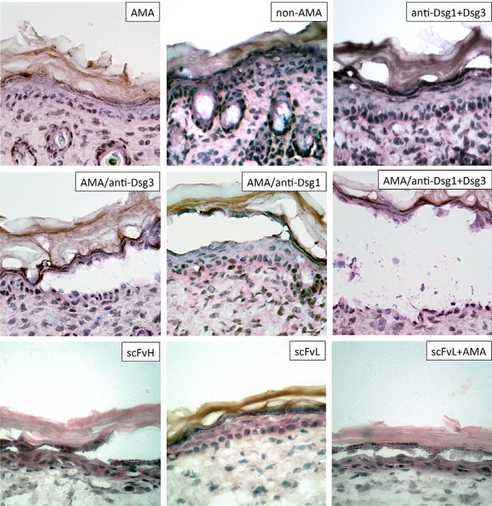
FIGURE 5.
Synergistic acantholytic activities of AMA and anti-Dsg antibodies in the neonatal mouse skin explant model. Representative images of epidermis in ∼3 × 3-mm pieces of truncal skin of neonatal BALB/c mice incubated at 37 °C and 5% CO2 for 24 h in PBS containing AMA (i.e. PVIgGs preabsorbed with a mixture of the cytosolic and cell membrane proteins of KCs), polyclonal rabbit anti-Dsg1, and/or goat anti-Dsg3 antibodies known to react with the respective mouse Dsg molecules or high (650 ng/μl; scFvH) versus low (163 ng/μl; scFvL) concentrations of scFv. Each experiment was repeated at least three times. Magnification = ×40.

