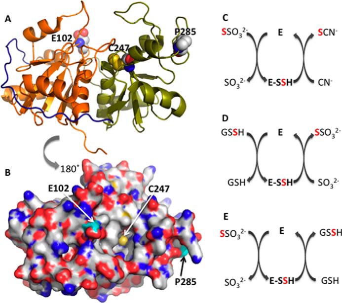
FIGURE 1.

Structure and reactions catalyzed by rhodanese. A and B, crystal structure of bovine rhodanese (Protein Data Bank code 1BOH). The N- and C-terminal domains are in orange and green, respectively, whereas the interdomain loop is in navy blue. The active site cysteine, Cys-247, and the polymorphic loci, Asp-102 and Pro-285, are shown in sphere representation. C–E, representative reactions catalyzed by rhodanese. In C and E, thiosulfate is the sulfane sulfur donor, whereas in D, GSSH is the sulfur donor. In each case, an enzyme-bound persulfide intermediate (E-SSH) is formed. Cyanide, sulfite, and GSH are the sulfur acceptors in C–E, respectively.
