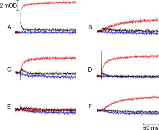
FIGURE 4.

Flash-activated heme b reduction. The traces were recorded for WT (A), G167P (B), 1Ala (C), G167P/1Ala (D), 2Ala (E), and G167P/2Ala (F) at pH 7 and an ambient potential of 100 mV. Kinetic transients at 560–570 nm were recorded without inhibitors (black lines) and in the presence of antimycin or myxothiazol (red or blue lines, respectively). mOD, milli-optical density units.
