FIGURE 6.
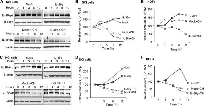
Effects of IL-36 induction on IL-1Rrp2 and IL-1RAcp accumulation. A, Western blot analysis examining the accumulation of IL-1Rrp2 in NCI cells treated with IL-36g and/or cycloheximide. The cells were collected at the indicated time after mock treatment or treatment with IL-36γ. β-actin accumulation served as a loading control in all samples. Cycloheximide (CH) was added to the cells to inhibit new rounds of translation to allow an estimate of the half-life of IL-1Rrp2 without additional protein synthesis. B, the half-life of IL-1Rrp2 after normalization to β-actin. NCI cells after mock treatment or treatment with IL-36γ or IL-1β in the presence or absence of cycloheximide over a 12-h time course. C, accumulation of IL-1RAcp increased over time and was not degraded rapidly after agonist addition. D, the half-life of IL-1RAcp in the presence of IL-36γ or cycloheximide in NCI cells. E, accumulation of IL-1Rrp2 in human dermal fibroblasts after mock treatment or treatment with IL-36γ. F, accumulation of IL-1RAcp in human dermal fibroblasts after mock treatment or treatment with IL-36γ. The data in B and D–F represent mean ± S.D. of three independent samples per time point.

