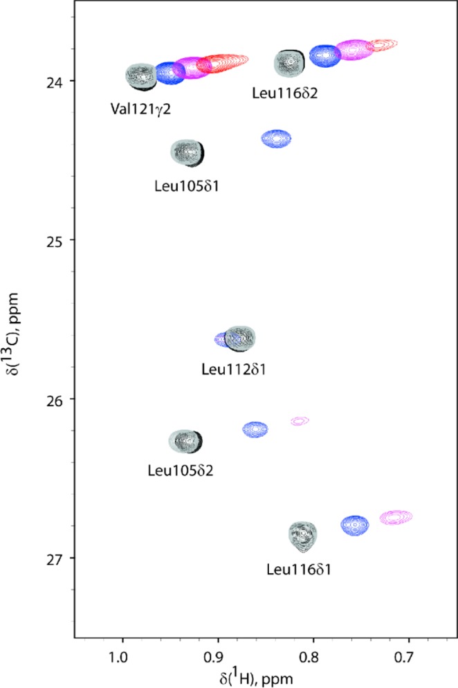
Figure 3.

Overlay of 2D NMR 1H–13C HSQC spectra of calmodulin C-lobe titrated with the lanthanoid La3+/Dy3+ complexed sevoflurane analogues. 1H–13C HSQC spectra of 0.1 mM calmodulin C-lobe (black) titrated with 0.05 mM (blue), 0.1 mM (purple), and 0.2 mM (red) Dy3+ complexed sevoflurane analogue are shown. Resonances transverse along approximately parallel lines with the addition of the paramagnetic ligand. Some peaks are broadened beyond detection at the higher concentration of the Dy3+ complexed sevoflurane analogue. The true δPCS are calculated with respect to the chemical shifts detected upon titration with 0.1 mM La3+ complexed sevoflurane analogue (gray). The sensitivity of the methodology is illustrated by the size of the induced pseudocontact shift (chemical shift differences for calmodulin in the presence of 0.1 mM Dy3+ or La3+ complexed sevoflurane analogue, spectra in magenta and gray, respectively) as compared to the size of the chemical shift changes induced by the nonparamagnetic ligand (chemical shift difference for calmodulin in the absence or presence of 0.1 mM La3+ complexed sevoflurane analogue, spectra in black and gray, respectively).
