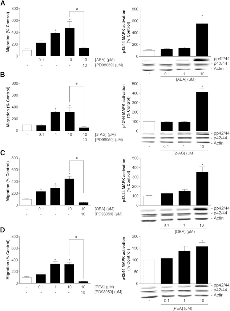
Fig. 4.
Impact of endocannabinoids and endocannabinoid-like substances on migration and p42/44 MAPK activation in MSCs. A–D (left panels): Concentration-dependent effects of AEA (A), 2-AG (B), OEA (C), and PEA (D) on migration of MSCs (Boyden chamber assays) following a 6 h incubation period with vehicle or the indicated concentration of test substances and impact of the p42/44 MAPK activation inhibitor PD98059 (1 h pretreatment) on the promigratory action of 10 μM of the respective test compound. A–D (right panels): Concentration-dependent effects of AEA (A), 2-AG (B), OEA (C), and PEA (D) on p42/44 MAPK phosphorylation in MSCs (Western blots) following a 2 h incubation period with vehicle or the indicated concentrations of test substances. β-actin was used as loading control. Histograms above the blots indicate activation of p42/44 MAPK determined by densitometric analyses of phosphorylated p42/44 MAPK normalized to that of nonphosphorylated p42/44. Percent control represents mean ± SEM compared with vehicle controls (100%) of n = 3 experiments using cells from one donor [left panels (A–D); right panel (B)] or n = 6 experiments using cells from two donors [right panels (A, C, D)]. *P < 0.05 versus vehicle; #P < 0.05 as indicated, one-way ANOVA plus post hoc Bonferroni (left panels) or Dunnett test (right panels).

