Fig. 7.
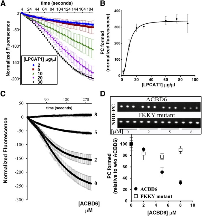
Effect of ACBD6 on the acyl-CoA transfer to lysoPC by LPCAT1. Real-time measurements of the incorporation of 16-NBD-C16:0-CoA into lysoPC were performed with 20 µM lysoPC and with 2 µM 16-NBD-C16:0-CoA as described in legend of Fig. 6. After 5 min of recording, the reactions (200 µl) were transferred into 750 µl of CHCl3/methanol (1:2), and lipids were extracted and NBD-PC was quantified as described in the Materials and Methods. A: Reactions were performed with increasing concentration of LPCAT1 microsomes. B: The amount of PC formed was plotted as a function of the concentration of LPCAT1. C: Reactions were performed with 30 µg of LPCAT1 microsomes and the indicated concentration of ACBD6 (0 to 8 µM). D: The amount of PC formed was plotted as a function of the concentration of ACBD6 or the mutant ACBD6.FKKY-AAAA. Inset: UV shadow imaging of the separated NBD-PC molecules by TLC. Reactions were performed in triplicate. Error bars represent the standard deviations of at least three measurements.

